rv lv ratio radiopaedia | what is rv Lv ratio rv lv ratio radiopaedia Right heart strain can often occur as a result of pulmonary arterial hypertension (and its underlying causes such as massive pulmonary emboli). Patients with . See more Mājaslapa. www .dagda .lv. Dagda Vikikrātuvē. Dagda ir pilsēta Krāslavas novadā Latgalē. Atrodas Latgales augstienes austrumu nogāzē, pie Dagdas ezera un Narūtas upītes, 36 km ziemeļaustrumos no Krāslavas, 267 km no Rīgas. Agrākā Dagdas rajona (1950-1962) un Dagdas novada (2009-2021) centrs.
0 · what is rv strain
1 · what is rv Lv ratio
2 · rv to Lv ratio radiology
3 · rv strain on echo
4 · right ventricle to left ratio
5 · right heart strain with pe
6 · right heart strain on ekg
7 · normal rv Lv ratio
Order Dabmar Lighting 2 x 2.5W & 12V JC-LED 36 LEDs X Cover Step Light - Black, LV-LED635-B at Zoro.com. Great prices & free shipping on orders over $50 when you sign in or sign up for an account.
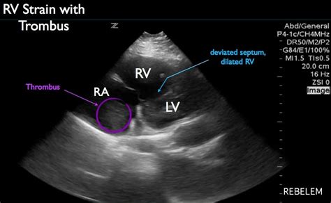
Right heart strain can often occur as a result of pulmonary arterial hypertension (and its underlying causes such as massive pulmonary emboli). Patients with . See moreThe reported sensitivity and specificity of CT in demonstrating right heart dysfunction are around 81% and 47% respectively 5. Described features include: 1. . See more
Right ventricular dysfunction usually results from either pressure overload, volume overload, or a combination. It occurs in a number of clinical scenarios, including: pressure .
Right ventricular enlargement (also known as right ventricular dilatation (RVD)) can be the result of a number of conditions, including: pulmonary valve stenosis. pulmonary arterial .Several methods to determine RV dysfunction on computed tomographic pulmonary angiography (CTPA) have been proposed. According to the latest European Society of Cardiology (ESC) . The right ventricular to left ventricular diameter (RV:LV) ratio measured at CT pulmonary angiogram (CTPA) has been shown to provide valuable information in patients with .
The most common measurements on CT scan were increased RV/LV ratio (21 studies), pulmonary artery measurements (6 studies), RV dilatation or increased size (5 .
RV volume—An increase in RV volume is one of the first signs of volume or pressure overload, or both (Figs. 4A, 4B, and 4C). It is most commonly and easily assessed by measuring the RV-to-LV short-axis ratio.
RV dilatation, as assessed by an elevated r ight-to-left vent ricular (RV/LV) diameter ratio in the four-chamber view, is associated with short-term mortality and is .The right ventricular to left ventricular diameter (RV:LV) ratio measured at CT pulmonary angiogram (CTPA) has been shown to provide valuable information in patients with pulmonary .
The primary aim of the study was to evaluate the accuracy of assessing the presence or absence of RV dilatation, defined as an RV/LV diameter ratio of ≥1.0, by three residents in internal . Right ventricular enlargement (also known as right ventricular dilatation (RVD)) can be the result of a number of conditions, including: pulmonary valve stenosis. pulmonary arterial hypertension. atrial septal defect (ASD) ventricular septal defect (VSD) tricuspid regurgitation. dilated cardiomyopathy. anomalous pulmonary venous drainage. Right ventricular dysfunction usually results from either pressure overload, volume overload, or a combination. It occurs in a number of clinical scenarios, including: pressure overload. cardiomyopathies: ischemic, congenital. valvular heart disease. arrhythmias. sepsis. It can manifest as right heart strain. Pre-capillary pulmonary hypertension is considered if the pulmonary artery wedge pressure (PAWP) is ≤15 mmHg, pulmonary vascular resistance (PVR) is ≥ 3 Wood units (WU) and mPAP is >20 mmHg. Post-capillary pulmonary hypertension is now defined as mPAP >20 mmHg and PAWP >15 mmHg.
what is rv strain
ratio non-compacted to compacted enddiastolic myocardium. affected left ventricular segments. left ventricular myocardial mass (compacted and non-compacted myocardium) left ventricular function. Treatment and prognosis. The only definitive treatment of left ventricular non-compaction is heart transplantation.
what is rv Lv ratio
RVD (right ventricular diameter): LVD (left ventricular diameter) ratio >1 on reconstructed four-chamber views. RVD:LVD ratio >1 on standard axial views is not considered to be a good predictor of right ventricular dysfunction 8Right heart strain evidenced by enlargement of the right ventricle (RV/LV ratio: 1.88) and reflux of contrast into the hepatic and azygous veins. No enlargement of the main pulmonary trunk. Subtle mosaic attenuation of the lungs in keeping with the multiple pulmonary emboli.There is mild cardiomegaly, primarily due to right-sided chamber enlargement, with straightening of the intraventricular septum and increase in the RV/LV ratio; associated reflux of IV contrast into the infrahepatic IVC and hepatic veins is present.Suggestion of right heart strain with an RV:LV ratio >1 and leftward deviation of the interventricular septum. Dependent atelectasis. Upper abdomen with extensive ascites.
Occlusive filling defects are identified on the low monoE recons in multiple segmental right lower lobe pulmonary arteries. No signs of right ventricular strain. RV/LV ratio is 0.8 (normal is <1.0). No dilation of the main pulmonary artery. The heart and mediastinal structures are normal. Reduced lung volumes with mild dependent atelectasis.
The parasternal long axis and apical four-chamber views on transthoracic echocardiography are often the primary views used to gain both a qualitative and quantitative appreciation of left ventricular enlargement. Features include 4: increased left ventricular internal end-diastolic diameter (LVIDd) Right ventricular enlargement (also known as right ventricular dilatation (RVD)) can be the result of a number of conditions, including: pulmonary valve stenosis. pulmonary arterial hypertension. atrial septal defect (ASD) ventricular septal defect (VSD) tricuspid regurgitation. dilated cardiomyopathy. anomalous pulmonary venous drainage.
Right ventricular dysfunction usually results from either pressure overload, volume overload, or a combination. It occurs in a number of clinical scenarios, including: pressure overload. cardiomyopathies: ischemic, congenital. valvular heart disease. arrhythmias. sepsis. It can manifest as right heart strain.
Pre-capillary pulmonary hypertension is considered if the pulmonary artery wedge pressure (PAWP) is ≤15 mmHg, pulmonary vascular resistance (PVR) is ≥ 3 Wood units (WU) and mPAP is >20 mmHg. Post-capillary pulmonary hypertension is now defined as mPAP >20 mmHg and PAWP >15 mmHg. ratio non-compacted to compacted enddiastolic myocardium. affected left ventricular segments. left ventricular myocardial mass (compacted and non-compacted myocardium) left ventricular function. Treatment and prognosis. The only definitive treatment of left ventricular non-compaction is heart transplantation. RVD (right ventricular diameter): LVD (left ventricular diameter) ratio >1 on reconstructed four-chamber views. RVD:LVD ratio >1 on standard axial views is not considered to be a good predictor of right ventricular dysfunction 8
Right heart strain evidenced by enlargement of the right ventricle (RV/LV ratio: 1.88) and reflux of contrast into the hepatic and azygous veins. No enlargement of the main pulmonary trunk. Subtle mosaic attenuation of the lungs in keeping with the multiple pulmonary emboli.There is mild cardiomegaly, primarily due to right-sided chamber enlargement, with straightening of the intraventricular septum and increase in the RV/LV ratio; associated reflux of IV contrast into the infrahepatic IVC and hepatic veins is present.
chanel rouge allure velvet stores

Suggestion of right heart strain with an RV:LV ratio >1 and leftward deviation of the interventricular septum. Dependent atelectasis. Upper abdomen with extensive ascites.
chanel rouge allure velvet emotive swatch
Occlusive filling defects are identified on the low monoE recons in multiple segmental right lower lobe pulmonary arteries. No signs of right ventricular strain. RV/LV ratio is 0.8 (normal is <1.0). No dilation of the main pulmonary artery. The heart and mediastinal structures are normal. Reduced lung volumes with mild dependent atelectasis.
rv to Lv ratio radiology
CV-Online is the place to find better career opportunities in all Baltic States - Latvia, Estonia, and Lithuania.
rv lv ratio radiopaedia|what is rv Lv ratio


