lv hypertrophy ecg | lv hypertrophy ecg criteria lv hypertrophy ecg Also known as: Left Atrial Enlargement (LAE), Left atrial hypertrophy (LAH), left . It’s the first “nice” mechanical watch for many enthusiasts, and for some, it’s the only watch they’ll ever own. Let’s discuss some key points to keep in mind if you’re considering purchasing an Omega Speedmaster. There’s even a slightly controversial one towards the end. 1. The Size of the Omega Speedmaster
0 · what is lvh on ecg
1 · signs of lvh on ecg
2 · lvh with repolarization abnormality ecg
3 · lv hypertrophy ecg criteria
4 · left ventricular hypertrophy with repolarization abnormality
5 · left ventricular hypertrophy life in the fast lane
6 · ecg showing lvh
7 · ecg in left ventricular hypertrophy
$5,666.00
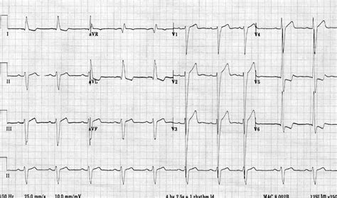
The left ventricle hypertrophies in response to pressure overload secondary to conditions such as aortic stenosis and hypertension. This results in increased R wave amplitude in the left-sided ECG leads (I, aVL and V4-6) and increased S wave depth in the right-sided .The ECG Made Practical 7e, 2019; Kühn P, Lang C, Wiesbauer F. ECG Mastery: .ECG Pearl. There are no universally accepted criteria for diagnosing RVH in .Also known as: Left Atrial Enlargement (LAE), Left atrial hypertrophy (LAH), left .
The delay between activation of the RV and LV produces the characteristic “M .The ECG Made Practical 7e, 2019; Kühn P, Lang C, Wiesbauer F. ECG Mastery: .
Kühn P, Lang C, Wiesbauer F. ECG Mastery: The Simplest Way to Learn the .Learn how to interpret ECG changes in LVH, such as large R-waves in left-sided leads and deep S-waves in right-sided leads. Find out the common causes, .
dior couture lipstick
The left ventricle hypertrophies in response to pressure overload secondary to conditions such as aortic stenosis and hypertension. This results in increased R wave .

The most common causes of left ventricular hypertrophy are aortic stenosis, aortic regurgitation, hypertension, cardiomyopathy and coarctation of the aorta. There are several ECG indexes, . Left ventricular hypertrophy changes the structure of the heart and how the heart works. The thickened left ventricle becomes weak and stiff. This prevents the lower left heart . Left ventricular hypertrophy (LVH) refers to an increase in the size of myocardial fibers in the main cardiac pumping chamber. Such hypertrophy is usually the response to a .
Electrocardiogram. Also called an ECG or EKG, this quick and painless test measures the electrical activity of the heart. During an ECG, sensors called electrodes are .
CONTENTS LAD (left axis deviation) LAHB (left anterior hemiblock) iLBBB (incomplete left bundle branch block) LVH (left ventricular hypertrophy) Diagnostic criteria . According to the American Society of Echocardiography and/European Association of Cardiovascular Imaging, LVH is defined as an increased left ventricular mass index (LVMI) .Left ventricular hypertrophy can be diagnosed on ECG with good specificity. When the myocardium is hypertrophied, there is a larger mass of myocardium for electrical activation to .
Interval between QRS and R-wave peak in V5 or V6 ≥ 0.05 second. 1. Sokolow-Lyon. V1 S wave + V5 or V6 R wave ≥ 35 mm. or. aVL R wave ≥ 11 mm. N/A. ECG = electrocardiography; LVH . Left ventricular hypertrophy with increased precordial voltages and non-specific ST segment and T-wave abnormalities. Deep, narrow (“dagger-like”) Q waves in lateral (I, aVL, . The left ventricle hypertrophies in response to pressure overload secondary to conditions such as aortic stenosis and hypertension. This results in increased R wave .
The most common causes of left ventricular hypertrophy are aortic stenosis, aortic regurgitation, hypertension, cardiomyopathy and coarctation of the aorta. There are several ECG indexes, .
Left ventricular hypertrophy changes the structure of the heart and how the heart works. The thickened left ventricle becomes weak and stiff. This prevents the lower left heart . Left ventricular hypertrophy (LVH) refers to an increase in the size of myocardial fibers in the main cardiac pumping chamber. Such hypertrophy is usually the response to a .
Electrocardiogram. Also called an ECG or EKG, this quick and painless test measures the electrical activity of the heart. During an ECG, sensors called electrodes are . CONTENTS LAD (left axis deviation) LAHB (left anterior hemiblock) iLBBB (incomplete left bundle branch block) LVH (left ventricular hypertrophy) Diagnostic criteria .
According to the American Society of Echocardiography and/European Association of Cardiovascular Imaging, LVH is defined as an increased left ventricular mass index (LVMI) .Left ventricular hypertrophy can be diagnosed on ECG with good specificity. When the myocardium is hypertrophied, there is a larger mass of myocardium for electrical activation to .
what is lvh on ecg
Interval between QRS and R-wave peak in V5 or V6 ≥ 0.05 second. 1. Sokolow-Lyon. V1 S wave + V5 or V6 R wave ≥ 35 mm. or. aVL R wave ≥ 11 mm. N/A. ECG = electrocardiography; LVH .
dior lipstick 625
signs of lvh on ecg
lvh with repolarization abnormality ecg
Coordinates: 35°53′N 14°27′E. Malta is an island in Southern Europe. It is the largest and most populous of the three major islands that constitute the Maltese Archipelago .The .
lv hypertrophy ecg|lv hypertrophy ecg criteria


